
Scientific sessions
")
Seminarios CTB-MINA en Biomateriales y Neurotecnología - Curso 2024/25
Como parte de los eventos oficiales del programa “Madrid Innovative Neurotech Alliance” (https://mina-cm.es/), que reúne grupos de investigación en neurotecnología y biomateriales, con énfasis en la salud y su impacto social, desde el CTB organizamos sesiones semanales enfocadas en la formación de estudiantes de doctorado, máster y grado en biomateriales, bioingeniería y áreas afines. Anualmente se celebran entre 12 y 15 sesiones de 1,5 horas de duración, donde los estudiantes presentan el progreso de sus investigaciones de doctorado, TFM o TFG en los campos de biomateriales y bioingeniería. También investigadores del
CTB y MINA-CM participan en los seminarios, fomentando la interacción y el intercambio de ideas.
SEMINARIO: Mobile and wearable technologies for health research. Dario Salvi, PhD – Associate Professor at Malmö University
Abstract: Thanks to their embedded sensors and connectivity features, ubiquitous smartphones and wearable devices offer an excellent opportunity for health research, including distributed recruitment, data collection, and interventions. I am going to present Mobistudy, a mobile-based platform for health research and results from a number of studies that have been conducted using it. I will also discuss challenges we have faced during this research including technical constraints, user aspects, legal and data protection issues.
Biosketch of the speaker: Dario Salvi holds an MsC in Telecommunication Engineering (Naples, Italy) and a PhD in Biomedical Engineering (Madrid, Spain). His research interests have focussed on the use of IT systems in healthcare, particularly in home monitoring, tele-health and clinical applications for consumer technologies such as smartphones and wearable devices. He is currently Associate Professor in Computer Science in Sweden where he works in a group that does research on digital health and collaborates with academics and clinicians in several countries to support research through apps and systems he develops.
- Miércoles, 13 de febrero de 2025 – 12:00 Salón de Actos del Centro de Tecnología Biomédica (UPM) Campus de Montegancedo, Pozuelo de Alarcón (Madrid)
Seminarios CTB-MINA en Biomateriales y Neurotecnología - Curso 2023/24
Como parte de los eventos oficiales del programa “Madrid Innovative Neurotech Alliance” (https://mina-cm.es/), que reúne grupos de investigación en neurotecnología y biomateriales, con énfasis en la salud y su impacto social, desde el CTB organizamos sesiones semanales enfocadas en la formación de estudiantes de doctorado, máster y grado en biomateriales, bioingeniería y áreas afines. Anualmente se celebran entre 12 y 15 sesiones de 1,5 horas de duración, donde los estudiantes presentan el progreso de sus investigaciones de doctorado, TFM o TFG en los campos de biomateriales y bioingeniería. También investigadores del
CTB y MINA-CM participan en los seminarios, fomentando la interacción y el intercambio de ideas.
Presentación del Premio de la Cátedra Cajal, otorgado a la investigadora del laboratorio de Biomateriales del CTB, la Dra. Nahla Jemni.
En el mismo intervendrá D. Santiago Ramón y Cajal Agüeras, presidente del comité científico de la Cátedra Cajal de la Universidad de Zaragoza, sobrino biznieto del premio Nobel y catedrático de Anatomía Patológica de la Universidad Autónoma de Barcelona y académico de número de la Real Academia Nacional de Medicina de España.
Además, contaremos con la presencia de Dª. Carmen González Madrid, presidenta de la comisión mixta de la Cátedra Cajal y de la Fundación Merck Salud y de D. José Luis Rodríguez Peralto, presidente de la Sociedad Española de Anatomía Patológica, Jefe de Servicio de Anatomía Patológica del Hospital Universitario 12 de Octubre, en Madrid y Catedrático de Anatomía Patológica de la Universidad Complutense de Madrid (UCM). Nos acompañará también Dª. Asunción Gómez-Pérez, Vicerrectora de Investigación, Innovación y Doctorado de la UPM.
- 21 de abril a las 11:00 horas.
- Inscripciones: https://eventos.upm.es/98091/programme/presentacion-del-premio-de-la-catedra-cajal-de-la-universidad-de-zaragoza.html
Seminarios CTB-MINA en Biomateriales y Neurotecnología - Curso 2022/23
Como parte de los eventos oficiales del programa “Madrid Innovative Neurotech Alliance” (https://mina-cm.es/), que reúne grupos de investigación en neurotecnología y biomateriales, con énfasis en la salud y su impacto social, desde el CTB organizamos sesiones semanales enfocadas en la formación de estudiantes de doctorado, máster y grado en biomateriales, bioingeniería y áreas afines. Anualmente se celebran entre 12 y 15 sesiones de 1,5 horas de duración, donde los estudiantes presentan el progreso de sus investigaciones de doctorado, TFM o TFG en los campos de biomateriales y bioingeniería. También investigadores del
CTB y MINA-CM participan en los seminarios, fomentando la interacción y el intercambio de ideas.
Nueva edición de la Competición actúaupm. Organizada por el CAIT (Centro de Apoyo a la Innovación Tecnológica) de la UPM
La inscripción está abierta hasta el 21 de marzo de 2023
¿En algún momento te has planteado emprender en la Universidad?, ¿te gustaría conocer el potencial de tu tecnología y aprender cómo sacarle el mayor partido?, ¿quieres impactar en el mercado y sociedad con tu investigación?
Desde Emprendimiento UPM te presentan una nueva edición de la Competición actúaupm, que este año cumple 20 ediciones, y viene cargada de sorpresas. Una oportunidad única para conocer el potencial de tu tecnología con la ayuda de grandes profesionales, que llevan más de dos décadas dedicándose a ello.
¡Así que no dejes pasar la oportunidad de inscribirte en la 20ª Competición actúaupm! Solo tienes que presentar tu idea o ideas antes del 21 de marzo.
Y, ¿qué te llevas si participas?
- Formación avanzada en emprendimiento
• Asesoramiento por parte del equipo de emprendimiento de la UPM y de tutores expertos
• Participación en sesiones, talleres y diferentes actividades orientadas al desarrollo de tu idea
• 45.000 euros en premios
• Feedback constante para la evolución del proyecto
• Networking con el resto de participantes y profesionales del sector
• Interesante red de contactos gracias a los 20 años de experiencia del programa
• Garantía de confidencialidad
• Acceso al Club Alumni
• Apoyo en la difusión y en la búsqueda de financiación
• … ¡Y mucho má
- Más info: www.upm.es/actuaupm
- Inscripciones: https://www.upm.es/S2i/fdin/index.jsp?alias=Formulario20actuaupm
"Lab-in-a-cell analysis enabled by SPAchip® Technology"
Seminario impartido por Antonio Quílez-Álvarez, PhD – Head of Applications at A4cell.
Abstract At A4Cell we develop lab-in-a-cell devices for living single-cell analysis of intracellular parameters. These SPAchip® devices (Suspended-Planar Arrays) are based on inert silicon oxide, on which multiple fluorescent probes can be deposited to provide intracellular readouts over long culture periods. The SPAchip® is internalized into the cell without affecting cell viability or proliferation capabilities. A4Cell proposes Cytocheck SPAchip® detection kits as cellomic reagents to measure intracellular analytes by fluorescence, they are compatible with fluorescence microscopy and flow cytometry. Its readouts can be included in cell-based assays for drug discovery and target identification in high throughput mode. In this talk we will cover the main aspects of this novel technology and the main applications we have developed to maximize assay relevance with intracellular data of live cells.
Biosketch of the speaker:
Dr. Antonio Quílez-Álvarez is Head of Applications at A4Cell. He graduated as a Materials Engineer from the Technical University of Madrid (UPM), Spain, where he obtained a collaboration grant from the Ministry of Education for an internship at Pharmamar (Spain). M.Sc. in Nanobiotechnology at University of Aalborg, Denmark, he registered a patent in biomarker assay development while working at Nordic Bioscience (Denmark). He was a Marie Curie predoctoral fellow at the National Center for Cardiovascular Research (CNIC), Spain, where he actively participated in different projects in the fields of Mechanobiology, assay development and High-Content Screening of cancer cellular models leading to publications in international journals and the obtention of his PhD from the Autonomous University of Madrid (UAM), Spain. He has given oral presentations of his work in international conferences and participated in multiple scientific ourteach activities.
- 8 Febrero 2023 – 11:00h
- Salón de Actos del Centro de Tecnología Biomédica. Campus de Montegancedo s/n. Pozuelo de Alarcón (Madrid)
"The end of medicine as we know it - And why our health has a future"
Charla impartida por Dr. Harald Schmidt
Resumen We do not understand the causality of almost any human disease and thus declare them as chronic and complex. Instead of treating causes we must treat symptoms, which is highly ineffective. For so-called complex diseases, most drugs are highly ineffective, and the success rate of drug discovery is in a constant decline. To this, reproducibility issues with almost 60% of all biomedical publications and wrong incentives such as the obsession with high impact factor papers contribute. The biggest conceptual error in Medicine was to split up the human body organ by organ, including organ-specific research disciplines and to pretend one can define a disease within one organ. Systems Medicine overcomes this roadblock by re-integrating the human body in an evidence-based manner enabled by data science and bioinformatics. Systems medicine’s therapeutic arm, network pharmacology, revolutionizes how we define, diagnose, treat, and ideally cure diseases. Descriptive disease phenotypes are replaced by mechanistic endotypes defined by causal, multi-target signaling modules that also explain respective comorbidities. These modules are however distinct from classical pathways, which we now recognize to be not more than highly curated mind maps of signaling events. Such modules often contain several fragments of several different canonical pathways. Precise and effective therapeutic intervention is subsequently achieved by synergistic multi-compound network pharmacology, ideally through drug repurposing, obviating the need for drug discovery, and speeding up the clinical translation. Network pharmacology is, however, not to be confused with the current combination therapies with multiple non-synergistic drugs targeting non-causal proteins that rather treat symptoms but do not cure disease. Eventually, how we conduct biomedical research, define signalling, diagnose and treat diseases, and practice medicine and will radically change.
- 20 Septiembre de 2022
- Salón de Actos del Centro de Tecnología Biomédica. Campus de Montegancedo s/n. Pozuelo de Alarcón (Madrid)
"Training-on-a-Chip: a multi-organ device to study the effect of muscle exercise on insulin secretion in vitro"
Charla impartida por Dr. Javier Ramón Azcón
Resumen Unraveling the mechanisms leading to type 2 diabetes (T2D) is fundamental in searching for new molecular drugs to prevent and control this disease. Organ-on-a-chip (OOC) devices offer new T2D disease modeling and drug discovery approaches by providing biologically relevant models of tissues and organs in vitro integrated with biosensors. Miniaturized biosensors systems and advanced tissue fabrication procedures enabled researchers to create multiple tissues on a chip with a high degree of control over experimental variables for high-content screening applications. In this work, we have fabricated a biomimetic multi-OOC integrated device composed of skeletal muscle and pancreatic cells to study the impact of exercise on insulin secretion. Both engineered tissues are integrated with optical biosensing technology to monitor contraction-induced myokine secretion and their effect on beta-cells insulin production (fig. 1). Our cross-talk platform would improve drug test approaches and provide a new model to study the loss of pancreas functionality associated with T2D.
- 12 Septiembre de 2022
- Salón de Actos del Centro de Tecnología Biomédica. Campus de Montegancedo s/n. Pozuelo de Alarcón (Madrid)
"Specific immunoglobulins against sars-cov-2 in human samples and its validation with the elisa technique"
NanoSpain 2022
A.M.M. Murillo 1,2, , J. Tomé-Amat 3, Y. Ramírez 1,2, M. Garrido-Arandia 3, L.G. Valle 1,4, G. HernandezRamírez 3, L. Tramarin 1,4,5, P. Herreros 1,4,5, B. Santamaría 1,4,6, A. Díaz-Perales 3,4, M. Holgado 3,4,5
Resumen Severe acute respiratory syndrome coronavirus 2 (SARS-CoV-2) is an RNA virus responsible for the pandemic that started in March 2020 and caused over 2.1 million deaths worldwide. Therefore, it is necessary to test the majority of the population in a fast, cheap, easily, and reliable manner. In this work we report the development of a novel in vitro diagnostic system based on biophotonic sensing cells (BICELLs) (figure 1) [1-3] to detect specific immunoglobulins in serum and saliva against SARS-CoV-2. This system aims the label-free detection of anti-SARS-CoV-2 IgG, IgM, and IgA of patients testing PCR-positive (figure 1), using the interferometric optical detection method (IODM). The results obtained were correlated and validated with those obtained by ELISA observing a significant correlation between the two techniques.
- May 17-20, 2022 Madrid
"La Pandemia por SARS-CoV-2 desde el Laboratorio Regional de Salud Pública"
Seminario impartido por el Prof. Pablo Barreiro García, Profesor titular en la Facultad de Ciencias de la Salud de la Universidad Europea de Madrid.
Resumen La infección por SARS-CoV-2 ha constituido unos de los mayores retos para la Medicina, que ha obligado también a adoptar medidas desde otros ámbitos: político, económico, social, familiar, etc. Desde el Laboratorio Regional de Salud Pública se han llevado a cabo diversos estudios con el fin de aportar evidencias científicas que guíen las políticas en materia de prevención. Los centros sociosanitarios fueron desde el inicio de la pandemia un foco de casos, donde además se dieron altas tasas de mortalidad. Acabada la primera ola pandémica, se llevó a cabo un estudio de seroprevalencia en la mayoría de los centros sociosanitarios de la Comunidad de Madrid, para identificar aquellas residencias que tenían mayor riesgo de sufrir nuevos brotes en el futuro. Posteriormente se hizo un seguimiento de los brotes que finalmente se dieron en estos centros y se establecieron los factores de riesgo para la aparición de nuevos casos. Otros colectivos de riesgo estudiados fueron la población dependiente que acude a centros de día, alumnos y profesores en centros educativos, funcionarios de prisiones y de fuerzas de orden público. El Laboratorio Regional de Salud Pública ha sido equipado con analizadores para el diagnóstico de la infección y para determinar la respuesta inmune, tanto humoral como celular, contra el SARS-CoV-2 o por la vacuna. Se han realizado varios estudios para validar la aplicación clínica y epidemiológica de estas técnicas. En esta conferencia se revisarán los principales resultados de estos estudios y cómo estos hallazgos han servido para guiar algunas decisiones de Salud Pública tomadas en la Comunidad de Madrid para afrontar las diversas olas pandémicas.
- 16 Febrero 2022 – 11:00h
- Salón de Actos del Centro de Tecnología Biomédica. Campus de Montegancedo s/n. Pozuelo de Alarcón (Madrid)
"Nanoactuators for Therapy and Diagnosis"
Seminario impartido por el Dr. Jesús Martínez de la Fuente, investigador del Instituto de Ciencia de Materiales de Aragón (ICMA-CSIC).
Este seminario se enmarca en el proyecto Neurocentro-CM (S2017/BMD-3760), financiado por la Comunidad de Madrid y el Fondo Social Europeo, y en la Comunidad Temática de Infraestructuras para la Salud, financiada por la Universidad Politécnica de Madrid.
Abstract In the last decades, inorganic nanoparticles have been steadily gaining more attention from a wide variety of fields as material science, engineering, physics, or chemistry. The very different properties compared to that of the respective bulk, and thus intriguing characteristics of materials in the nanometer scale, have driven nanoscience to be the center of many basic and applied research topics. Moreover, a wide variety of recently developed methodologies for their surface functionalization provide these materials with very specific properties such as drug delivery and circulating cancer biomarkers detection.
In this talk we describe the synthesis and functionalization of magnetic and gold nanoparticles as therapeutic and diagnosis tools against cancer. Gold nanoprisms (NPRs) have been functionalized with PEG, glucose, cell penetrating peptides, antibodies and/or fluorescent dyes, aiming to enhance NPRs stability, cellular uptake, and imaging capabilities, respectively.
- 24 Noviembre 2021 – 11:00h
- Salón de Actos del Centro de Tecnología Biomédica. Campus de Montegancedo s/n. Pozuelo de Alarcón (Madrid)
“Microgeles como entorno 3D en cultivos celulares en Ingeniería Tisular”
Conferencia del Dr. Jose Luis Gómez Ribelles, del Centro de Biomateriales e Ingeniería Tisular de la Universidad Politécnica de Valencia.
Resumen Llamamos microgel a una dispersión de microesferas en un medio acuoso . Cada partícula puede tener propiedades de gel en sí misma pero aunque no sea así el conjunto puede tener la consistencia de un hidrogel. La síntesis de las microesferas permite mimetizar la composición del nicho biológico, injertando bio moléculas de la matriz extracelular del tejido de interés en la superficie de las partículas.
La fracción en volumen de microesferas respecto al volumen total permite aplicarlos como entorno tridimensional en el cultivo celular de distintos tipos celulares. Altas proporciones de partículas hacen que las células estén rodeadas de material como si estuvieran encapsuladas en un hidrogel, pero con la diferencia de que las células tienen movilidad y pueden modificar su entorno para dar cabida a la matriz extracelular que producen y organizar el tejido neoformado. Por el contrario, bajas fracciones de microesferas permiten simular el entorno de células no adherentes que se cultivan en suspensión, pero poniendo a su alcance los constituyentes del entorno natural injertadas en las propias microesferas.
Son ejemplos de aplicación el cultivo de 3D de condrocitos y células madre mesenquimales, la regeneración del cartílago articular en un modelo animal y el cultivo de células plasmáticas tumorales como modelo de enfermedad del Mieloma Múltiple.
- 22 Noviembre 2021 – 11:00h
- Salón de Actos del Centro de Tecnología Biomédica. Campus de Montegancedo s/n. Pozuelo de Alarcón (Madrid)
“Development of biomimetic models of intestinal tissue: guiding cellular self-organization through biofabrication techniques”
Seminario impartido por la Dra. Elena Martínez Fraiz, líder del grupo de Biomimetic Systems for Cell Engineering del Institute for Bioengineering of Catalonia (IBEC).
Summary Epithelial tissues contain three-dimensional (3D) microstructures that guide cell self-organization at the tissue level. In the small intestine, crypts and finger-like villi microstructures improve its absorbance function, provides specific microenvironments and compartmentalizes cell types [1–3]. Despite its physiological relevance, tissue architecture and multicellular population are neglected in the standard in vitro models, thus compromising their predictive capabilities [4]. Our efforts in addressing these shortcomings by including key elements to mimic the native tissue in vitro will be discussed in this talk. First, this will include strategies to promote cell’s self-organization capabilities giving rise to crypt-villus domains on 2D monolayers [5], and strategies to engineer cell spatial positioning through micropatterning. Then, our approach to include the 3D architecture of the tissue will be addressed. In here, light-based biofabrication techniques to produce 3D villus-like structures [6,7] will be discussed. Finally, I will introduce our biofabrication proposal to produce tissue engineered models that include the epithelial and the stromal compartments [8]. Improving the prediction capabilities of cell-based assays is a growing strategy to lead to more efficient drug development processes. As 2D-based systems are showing their limits, new 3D strategies are gaining acceptance among the scientific community [9]. Our approaches aim to further accelerate this trend by providing feasible strategies to routinely incorporate 3D multicellular structures at the tissue level in cell culture systems.
- 18 Noviembre 2021 – 11:00h
- Salón de Actos del Centro de Tecnología Biomédica. Campus de Montegancedo s/n. Pozuelo de Alarcón (Madrid)
“Interpreting the mechanisms and meaning of MEG/EEG signals with the human neocortical neurosolver software”
Seminario impartido por la profesora Prof. Stephanie Jones Human Neocortical Neurosolver, Department of Neuroscience, Brown University, Providence RI, USA
Abstract Over the last decade, there has been a growing interest in understanding the brain mechanisms underlying the loss of brain homeostasis in the continuum of Alzheimer’s Disease (AD). Animal models suggest that the substrate for this phenomenon is the loss of the excitatory/inhibitory (E/I) balance due to the toxic effects of amyloid oligomers and plaques on inhibitory terminals. This hyperexcitability is presumed to underlie the observed increases in power and inter-area synchronization of alpha/beta frequency oscillations measured in humans with electro- and magneto-encephalography (M/EEG). While animal and human studies are highly synergistic, it is unknown if the hyperexcitability found in animal models is the origin of the hypersynchronization found in human neurophysiology. To bridge this gap, the current project will apply a recently developed computational neural modeling framework uniquely designed to link human macroscale M/EEG signals to the underlying cellular and circuit level dynamics that can be interrogated with invasive animal recordings or other imaging modalities, namely Human Neocortical Neurosolver (HNN) (https://hnn.brown.edu).
About Prof. Stephanie Jones Her research integrates human brain imaging and computational neuroscience methods to study brain dynamics in health and disease. She works closely with animal neurophysiologist and clinicians to develop data constrained neural models that are functionally and translationally relevant. The main theme of her lab’s research is to develop biophysically principled models of neural circuits that bridge the critical gap between human brain imaging signals (MEG/EEG) and their underlying cellular and network-level generators. Current projects apply such interdisciplinary techniques to study the mechanisms and functions of neural dynamics, including brain rhythms, in healthy functions such as perception, attention, and decision making, and in neural pathologies such as Parkinson’s disease and Essential Tremor. They also study the impact of brain stimulation (DBS, tACS, TMS), and mind-body practices, on brain dynamics with the ultimate goal of improving treatments for neuropathology.
- 16 Noviembre 2021 – 11:00h
- Salón de Actos del Centro de Tecnología Biomédica. Campus de Montegancedo s/n. Pozuelo de Alarcón (Madrid)
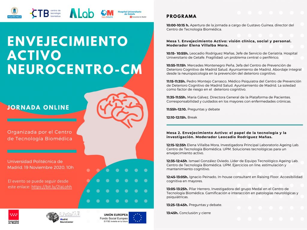
Seminario online de envejecimiento activo
- 19 noviembre 2020 Online
Seminario online de seminario Mecanobiología
- 21 Octubre 2020

The morphospace of language networks
Luíño Seoane – Department of Physics, Massachusetts Institute of Technology, USALanguage can be described using networks of (semanticaly, syntacticaly, …) interacting objects. Besides, a general information theory approach incorporates a speaker, a hearer, and a noisy channel. A key, common element in such approach is a confusion matrix encoding which words name each one of the existing objects, naturally introducing networks again. This also allows us to measure costs associated to communication across the channel for hearers and speakers. A rich literature on least-effort language exploring the optimality of communication codes developed from these methods. However, no systematic analysis of the underlying landscape of language graphs has been performed. We do this here, finding a rather complex and heterogeneous morphospace of language networks. We also derive a series of results relevant for the least-effort study of human language, largely in contradiction with theoretical speculations in the literature. These analysis are complemented, for the first time, with an empirical study of English words. Based on the WordNet database, we locate English vocabulary within the language networks morphospace. The outcome of this empirical analysis stresses the role of referential particles for efficient communication and to explore this morphospace.
Brief CV
Luíño Seoane studied Physics at the Universidade de Santiago de Compostela and Computational Neuroscience at the Technische Universität Berlin before completing his PhD in Complex Systems at the Pompeu Fabra University. There, under the supervision of Ricard Solé, he studied Multiobjective Optimization, its connections to Statistical Mechanics, and how optimization trade-offs lead to phase transitions and other phenomenology in complex networks, in linguistics, and in Darwinian evolution. His research in Neuroscience spans from Brain-Computer Interfaces to correlates of consciousness, while he is interested in developing research in Machine Learning and models of spiking neurons.
- 22 Junio 2018 – 11:00h
- Salón de Actos del Centro de Tecnología Biomédica. Campus de Montegancedo s/n. Pozuelo de Alarcón (Madrid)
Current state of telehealth projects for diabetes and cardiac telemonitoring for chronically ill patients
Dieter Hayn (www.ait.ac.at/profile/detail/Hayn-Dieter/) is Senior Scientist at the Digital Healthcare Information Systems group of the AIT Austrian Institute of Technology (www.ait.ac.at).
In his presentation, he will describe the current state of AIT’s telehealth projects for diabetes (DiabMemory) and cardiac (HerzMobil) telemonitoring for chronically ill patients. AIT’s telehealth projects are already running in routine care in Austria, financed within the Austrian healthcare system, and they have been pilots for Austria’s currently developing tele-health-service standards, which are intended to link telehealth to the Austrian Electronic Health Record ELGA.
Dieter Hayn will also present current results of their Predictive Analytics Toolset for Healthcare (PATH), which supports the rapid setup of predictive modelling solutions for various applications. Results from different applications of PATH will be discussed, including prediction of Delirium at hospital admission; blood transfusion needs prior surgery, re-admission at discharge, etc. A current application of PATH in a real-world clinical scenario at a hospital in Graz (Austria) will be presented, including visualization tools for plausibility checks of machine learning results.
- 20 Junio 2018 – 11:00h
- Salón de Actos del Centro de Tecnología Biomédica. Campus de Montegancedo s/n. Pozuelo de Alarcón (Madrid)
Production of recombinant silk protein and some applications
Seminario impartido por los profesores Anna Rising y Jan Johansson, del Instituto Karolinska, (Estocolmo, Suecia)
- 14 Junio 2018 – 11:00h
- Salón de Actos del Centro de Tecnología Biomédica. Campus de Montegancedo s/n. Pozuelo de Alarcón (Madrid)
Neurociencia aplicada a los recursos humanos de las empresas, lo último del siglo XXI
Ponencia organizada por Brainvestigations, empresa española que estudia las ondas cerebrales con un objetivo claro: aplicar y llevar el conocimiento científico al mundo de los negocios.
Ciencia, employer branding, aplicaciones neurocientíficas para validar políticas de recursos humanos, y cómo las técnicas de neuroimagen pueden decodificar la mente para aportar datos muy importantes que en estos momentos es más difícil obtener de otro modo, son otros de los temas que se pretenden abordar en esta jornada
En el encuentro tomarán parte Fernando Maestú, catedrático y director del Laboratorio Neurociencia Cognitiva del CTB e Ignacio Belinchón, consultor de Recursos Humanos y consejero de Brainvestigations.
Para asistir al evento es necesario completar el siguiente formulario: www.brainvestigations.com/contacto
Más información, gcomunicacion@brainvestigations.com
- 7 Junio 2018
- Salón de Actos del Centro de Tecnología Biomédica. Campus de Montegancedo s/n. Pozuelo de Alarcón (Madrid)
SEMINARS: Introduction to the study of Cortical Microanatomy
- 20 March 2018
- Laboratorio Cajal de Circuitos Corticales. Centro de Tecnología Biomédica. Campus de Montegancedo s/n. Pozuelo de Alarcón (Madrid)
- Agenda
Javier deFelipe: “Nuevas tecnologías para el estudio del cerebro: Human Brain Project”
- 01/03/2018
- Seminarios de Biomedicina 2018
- Elemento de lista
Christophe Letellier: “Assessing the observability of complex networks: a nonlinear theory”
Tenemos el placer de invitarles a la conferencia que tendrá lugar el día 02 de marzo a las 12:00 en el Aula 01 de la planta cero del Centro de Tecnología Biomédica, impartida por el investigador Christophe Letellier.
The observability of a complex system refers to the property of being able to infer its whole state by measuring the dynamics of a limited set of its variables. Since in practice, monitoring all the variables defining the system’s state is experimentally unfeasible or inefficient, it is of utmost importance to develop a methodological framework addressing the problem of targeting those variables yielding full observability. Despite several approaches have been proposed, most of them neglect the nonlinear nature typically exhibited by complex systems and/or do not provide the space reconstructed from the measured variables. On the one hand, since nonlinearities are often related to a lack of observability, linear approaches cannot properly address this problem. On the other hand, finding the appropriate combination of sensors (and time derivatives) spanning the reconstructed space is a very time demanding computational task for large dimensional systems. Here, we adopt a nonlinear symbolic approach taking into account the nature of the interactions among variables and analyze the distribution of the linear and nonlinear load of the variables in the symbolic Jacobian matrix of the system. By means of two easy-to-implement criteria we are able to successfully identify the minimal set of variables (and their time derivatives) candidate to be measured for completing the reconstructed space. Our predictions are in full agreement with the analytical solution and drastically reduce the search for candidate variables, thus providing a key step to observe and model natural and man made complex systems of large dimension [1]. Some explicit examples will be provided [2]. If I have time, I will end with an application of observability for a follow-up in oncology.
- 02/03/2018. 12:30
- Aula 01 de la planta cero del CTB
Sidharam P. Pujari: “Romantic Surface (funcionalization of biomolecules on surfaces)”
Tenemos el placer de invitarles a la conferencia que tendrá lugar el día 27 de octubre a las 12:30 en el Salón de Actos del Centro de Tecnología Biomédica, impartida por el investigador de la Universidad de Wageningen, Sidharam P. Pujari.
CV S.P.Pujari: he received a B.Sc. in Chemistry at the Shivaji University, India and an M.Sc. at the University of Pune, India. He worked at the National Chemical Laboratory in Pune, India, and at the National Taiwan University Science and Technology, Taipei before starting a PhD in Prof. Zuilhofs labs in Wageningen University on covalently bound fluorinated monolayers. Subsequently he was a postdoc at the Zuilhof labs, and visiting scholar at the University of Texas at Dallas with Prof. Y. J. Chabal. Currently, he works as research associate position in Wageningen in the fields of materials and surface chemistry. He has published 40 articles with 2 (eu & us) patents with h index = 15.
- 27/10/2017. 12:30
- Salón de Actos del Centro de Tecnología Biomédica. Campus de Montegancedo s/n. Pozuelo de Alarcón (Madrid)
Workshop: PAPHOS, your solution in the Life Science and Health digital transformation
Health prediction and treatment processes have become increasingly complex: involving huge amounts of data; with information coming from diverse sources; and the need of accessing to real-time data for decision-making. On the other hand, biocomputing has become a cornerstone of life sciences in the recent years, with high performance computing and analytics solutions as drivers of this digital transformation.
In this workshop, hosted by Atos Spain S.A. as leader of PAPHOS consortium, we will present PAPHOS platform offering, a holistic solution to address healthcare givers, clinical researchers and life science professionals’ problems for an effective, smooth and affordable translation of big data analytics to their practice.
- 18/10/2017. 11:30-16:30 (CET)
- Atos Business Technology and Innovation Centre (BTIC) at Paseo Doce Estrellas 2, Madrid (metro Feria de Madrid)
Profesor James Yang: “Digital Human Modeling and Simulation for Engineering and Biomechanics”
Tenemos el placer de invitarles a la conferencia que tendrá lugar el día 16 de octubre a las 10:00 en el Salón de Actos del Centro de Tecnología Biomédica, impartida por el profesor James Yang.
Abstract: Digital human modeling and simulation plays an important role in product design,prototyping, manufacturing, sports biomechanics, and other areas. It reduces the number of design iterations and increases the safety and design quality of products. In this talk, I will first briefly review the state of art of digital human models, then I will summarize research projects carried out in my research lab. The first area is in engineering by investigating the optimization based digital human models to assist design and engineering, slips and falls. Applications include driver accommodation study and special population posture and motion prediction. The second area is healthcare engineering and spine biomechanics.
- 16/10/2017. 10:00
- Salón de Actos del Centro de Tecnología Biomédica. Campus de Montegancedo s/n. Pozuelo de Alarcón (Madrid)
- Profesor James Yang